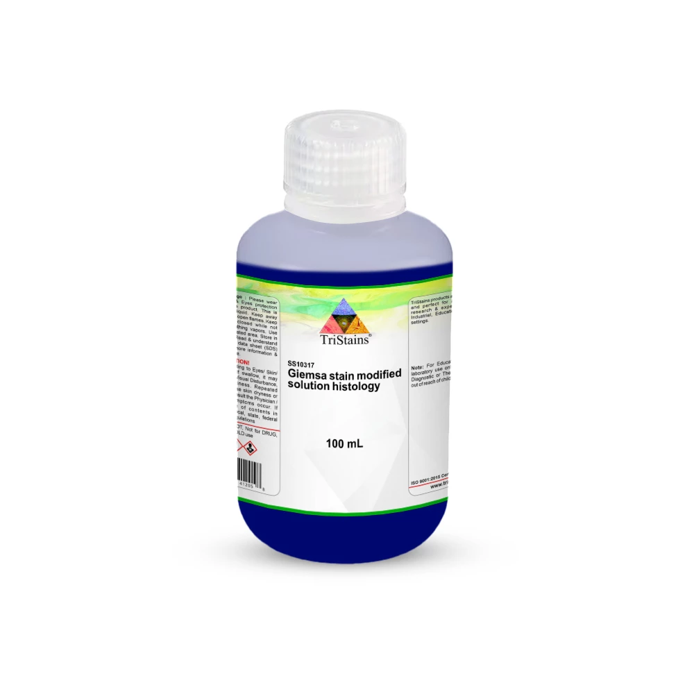
Giemsa Stain, Modified Solution-Histology, Buy Histological Stains solutions for Histology, Cytology, Microbiology, Hematology, Biology Lab from TriStains.
Giemsa Stain Modified Solution is a new version of the traditional Giemsa stain, which has been developed by means of methylene blue, eosin, and azure stains. Certain ratios of dyes and other materials in this altered formulation are well-tuned to improve its staining performance in special histological applications. This stain is becoming very popular in the field of histology, which is the scientific analysis of the microstructure of tissues, to provide a visualization of particular and accurate images.
Applications
- The stained Giemsa stain is highly used in histology as a stain to use in tissue sections. It is critical in the identification, discrimination, and examination of various tissue components and cellular structures made of dissimilar cell types to permit appropriate microscopic analysis.
Key features
- Reliable Staining: The staining formulation was designed to provide reliable, dependable, and reproducible staining results in routine and advanced histological analysis.
- Histologically Optimized: Optimized to be used in the histological technique and hence provide maximum color difference and structural clarity.
- Versatile Application: Extremely sensitive and can be used in diagnostic and research-based histological studies.
- Ready-to-Use Formulation: in a ready-to-use format to enhance efficiency of laboratories and ease the workflow.
General Description
The Giemsa Stain, Modified Solution-Histology is a more advanced version of the original Giemsa stain. It contains a mixture of methylene blue, eosin, and azure at specific proportions, along with other materials that enhance the quality of staining in histological tissue samples. This enhanced formula provides improved color separation, reduced background blurriness, and enhanced visualization of tissue structure and cellular elements in the microscopic view. It is particularly appreciated in diagnostic histology to assess cell morphology and the pathological alteration of tissue sections.
Want to Buy Giemsa Stain, Modified Solution - Histology? Buy high-quality Giemsa Stain, Modified Solution-Histology, and other high-quality TriStains reagents that are used in laboratories in histology, cytology, microbiology, hematology, and biology laboratories throughout the USA. TriStains products are known to be of quality and consistency, laboratory reliability, and longevity. Order Giemsa Stain, Modified Solution - Histology today for reliable and reproducible results from TriStains.
About Brand
TriStains provides a marketplace for histology and biological stains, which is comprehensive enough to encompass the peculiar requirements of laboratories specializing in Histology, Cytology, Microbiology, and Hematology. With a reputation for exceeding quality expectations, TriStains performance is outstanding which allows for resolution of cell and tissue components fundamental to life sciences to be clearly visualized. Each product under TriStains series is validated for accuracy, reliability and consistency. TriStains, which manufactures and markets stains and indicators in various packing, offers laboratories turn key solutions for all their staining and indicator needs, improving accuracy in every experiment.
Related Products


Redirect Notice
Tristain products are exclusively distributed by Dawn Scientific.
You are now being redirected to www.dawnscientific.com to complete your purchase.
Thank you.