Hematoxylin
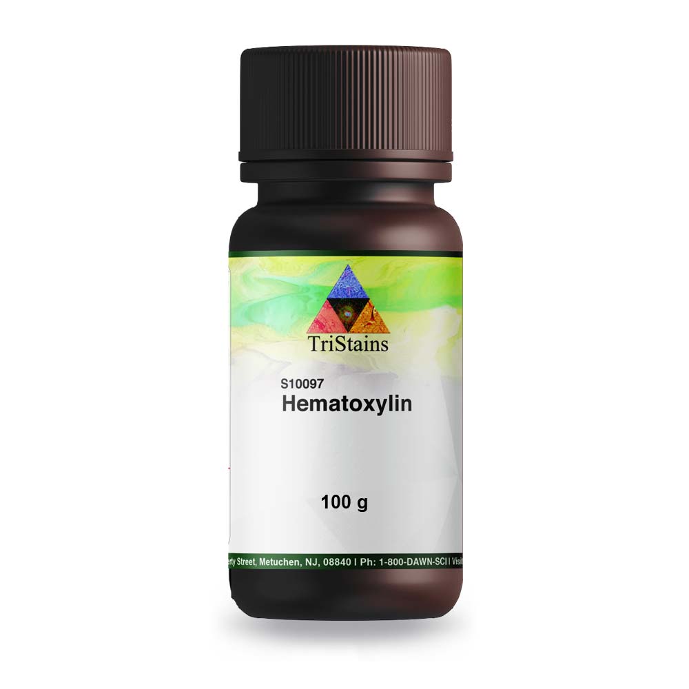
Hematoxylin is a natural dye extracted from the heartwood of certain trees, primarily from the logwood tree (Haematoxylum campechianum). It is commonly used as a biological stain in histology and pathology for highlighting cell nuclei. Tristains Biological Stains that offer range of stains used in Histology, Cytology, Microbiology and Hematology laboratories. Tristains meet the highest quality standards and give excellent color performance. Tristains series products are carefully tested to ensure accurate, reliable, and reproducible results. In this blog post, we are going to explore Hematoxylin’s description, extraction and purification, uses or applications and its formulations.
What is Hematoxylin?
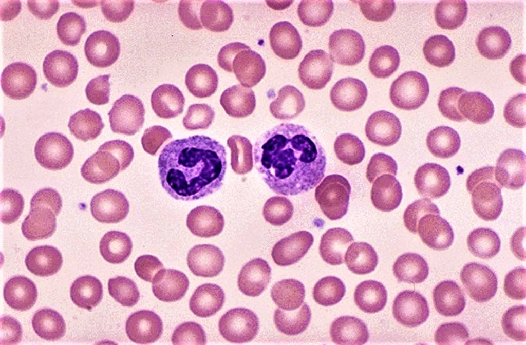
Hematoxylin, also known as Natural Black 1 or C.I. 75290, is derived from the wood of the logwood tree. Upon oxidation, it transforms into haematein, a compound displaying a vibrant blue-purple hue. This substance, in combination with a suitable mordant such as Fe (III) or Al (III) salts, is utilized to stain cell nuclei before microscopic examination. Cells that are stained with hematoxylin are referred to as basophilic.
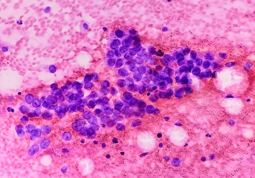
Hematoxylin is commonly paired with eosin, a counterstain, to enhance contrast in microscopy. The hematoxylin and eosin stain are widely used in histology and is considered a permanent stain, unlike temporary solutions like iodine in KI.
Another frequently used stain is phosphotungstic acid hematoxylin, which is a blend of hematoxylin and phosphotungstic acid.
Stain Formulations
Hematoxylin stain formulations are typically categorized based on the method of oxidation of hematoxylin and the choice of mordant. Hematoxylin stains can be naturally oxidized by exposure to air and sunlight, or chemically oxidized usingsodium iodate, which is more common in commercially prepared solutions.
Usually, only a sufficient amount of oxidizer is added to convert half of the hematoxylin to haematein, allowing the rest to oxidize naturally during use. This prolongs the useful life of the staining solution as more haematein is generated, with some haematein further oxidizing to oxyhaematein. Aluminum is the most commonly used metallic salt mordant, while other mordants include salts of iron, tungsten, molybdenum, and lead.
Extraction and Purification of Hematoxylin
Hematoxylin has been synthesized, but not in economically feasible quantities. In the past, hematoxylin was extracted in Europe from exported logwood, but now extraction occurs closer to where the logwood is harvested. Large-scale extraction is achieved through the ‘French process’ or the ‘American process’. The dye can be sold as a liquid or crystalline form. Modern production methods use water, ether, or alcohol as solvents. The commercial product can vary in impurities and hematoxylin to haematein ratio. This variability can affect the interaction with tissue samples. Certification by the Biological Stain Commission indicates performance in standardized tests, but not actual purity.
Uses of Hematoxylin
- Hematoxylin and eosin (H&E) staining is a widely utilized technique in histology, commonly employed in pathology labs to examine tissue samples. This staining method enables researchers and pathologists to observe the structure and morphology of cells and tissues, facilitating the diagnosis of different diseases.
- Hematoxylin is also a component of the Papanicolaou stain (or PAP stain) which is widely used in the study of cytology specimens, notably in the PAP test used to detect cervical cancer
- In the past, hematoxylin was extensively used to create blacks, blues, and purples on various textiles. It served as a crucial industrial dye until synthetic dyes became available as suitable alternatives. Nowadays, hematoxylin finds application in dyeing silk, leather, and sutures.
- Additionally, it has been utilized as the primary component in writing and drawing inks.
Aluminum Hematoxylin Solutions
There are two primary Alum Hematoxylin solutions commonly used: Ehrlich’s Hematoxylin and Harris Hematoxylin. These solutions provide a light transparent blue stain to the nucleus, which quickly changes to red when exposed to an acid.
- Ehrlich’s Hematoxylin is commonly employed for regressive staining, where it is differentiated using 1% HCl in 70% Acid-Alcohol until the nucleus is specifically stained. Mucopolysaccharide substances like cartilage and cement lines of bones are also deeply stained in a blue hue. The typical staining duration ranges from 15 to 40 minutes.
- Harris Hematoxylin is extensively utilized for standard nuclear staining in Exfoliative Cytology and for the staining of sex chromosomes. The typical duration for staining is between 5 and 20 minutes.
- Cole’s Hematoxylin is an additional Alum Hematoxylin solution that is suggested for regular use, particularly when used consecutively with Celestine Blue.
- Mayer’s Hematoxylin exhibits a higher level of activity compared to Ehrlich’s Solution, resulting in minimal or no staining of Mucopolysaccharides. This particular stain is employed in the Celestine Blue Hemalum technique for Nuclear Staining.
Iron Hematoxylin Solutions
Two main Iron Hematoxylin solutions are commonly used in labs: Weigert’s Solution (with Ferric Ammonium Chloride) and Heidenhain’s Solution (with Ferric Ammonium Sulfate). Both acts as oxidizing agents and should not be stored for long periods. These solutions can stain tissues fixed in various fixatives, even those stored in alcohol for a long time. The stained structures appear black or gray, reducing eyestrain and making it useful for photomicrography.
- Weigert’s Solution serves as the conventional Iron Hematoxylin utilized in laboratory settings, particularly for highlighting muscle fibers and connective tissues. It is highly advisable in cases where prior stains have acidic components (such as Van Gieson’s Stain with Picric Acid) that may fade nuclei stained with Alum Hematoxylin.
- Heidenhain’s Hematoxylin is a cytological stain that is highly recommended for the regressive staining of thin sections. It is commonly used to demonstrate nuclear and cytoplasmic inclusions, including chromatin, chromosomes, and mitochondria. Additionally, it effectively stains voluntary muscle striations and myelin.
Where To Buy
Buy premium quality Biological stain & Indicators from TriStains®. All TriStains® Products are exclusively distributed by Dawn Scientific Inc.
www.tristains.com
www.dawnscientific.com
About Tristains Brand
Biological Stains are a major part of all Histology, Hematology and Cytology labs and we understand that quality is of utmost importance, so try Premium quality biological stains TriStains® see the difference. TriStains® offers all types of stains and indicators in ready to use solutions as well as solid forms for more information please visit our website www.tristains.com or email us on [email protected] for any Query/information we will be happy to help you.
Disclaimer: All the information on this blog is published in good faith and for general information purpose only. The information contained in this Blog might be provided on an “as is” based on Wikipedia, Google and other scientific articles. We are not liable for any injuries or damages for the use of your information. Please do your own research before you use this information for any purpose.


